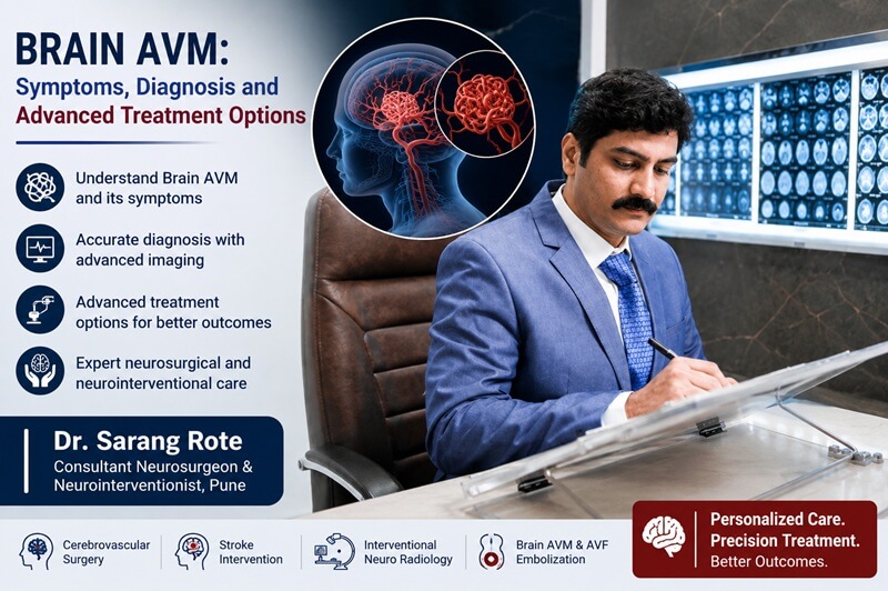
Brain AVM: Symptoms, Diagnosis and Advanced Treatment Options
A brain arteriovenous malformation (AVM) is an abnormal network of blood vessels in the brain in which arteries connect directly to veins without the normal capillary network in between.
Because blood flows differently through an AVM, the surrounding blood vessels and brain tissue can be placed under abnormal pressure. Some brain AVMs may cause no symptoms and may be discovered incidentally during brain imaging, while others can cause seizures, headaches, neurological symptoms or bleeding in the brain.
Early diagnosis and specialist evaluation are important because the appropriate management of a brain AVM depends on its size, location, blood-vessel anatomy, symptoms, previous bleeding and the patient's overall health.
What Is a Brain AVM?
A brain AVM, or cerebral arteriovenous malformation, is a tangle of abnormal blood vessels connecting arteries and veins in the brain.
Normally, blood travels from arteries into smaller vessels called capillaries before reaching the veins. Capillaries help regulate blood flow and allow oxygen to reach brain tissue.
In an AVM, blood passes directly from arteries to veins through abnormal vessels. This can create high-flow conditions and place stress on the blood vessels.
Some AVMs remain stable and may never cause symptoms. Others can bleed or affect surrounding brain tissue and lead to neurological problems.
What Causes a Brain AVM?
The exact cause of brain AVMs is not clearly known.
Many brain AVMs are believed to develop before birth during the formation of the brain's blood vessels. In rare cases, vascular malformations may be associated with inherited conditions.
A brain AVM is not usually caused by something a person has done during their lifetime.
Because AVMs can remain silent for many years, some people may not know they have one until symptoms develop or a brain scan is performed for another reason.
Brain AVM Symptoms
The symptoms of a brain AVM can vary depending on its size, location and whether bleeding has occurred.
Some people have no symptoms at all.
Possible symptoms include:
- Headaches
- Seizures
- Weakness in an arm or leg
- Numbness or altered sensation
- Problems with balance or coordination
- Vision problems
- Difficulty speaking
- Difficulty understanding language
- Changes in memory or thinking
- Dizziness
- Confusion
- Loss of consciousness in serious cases
Symptoms can differ considerably between patients because different areas of the brain control different functions.
Seizures and Brain AVM
Seizures are one of the possible symptoms of a brain AVM.
An AVM can irritate or affect nearby brain tissue, which may contribute to seizure activity.
A person who develops a new seizure, particularly without a previous history of seizures, should receive appropriate medical evaluation.
Brain imaging may be recommended to determine whether a structural abnormality such as an AVM is present.
Headache and Brain AVM
Headaches can occur in people with brain AVMs.
However, a headache alone does not necessarily mean that a person has an AVM because headaches have many possible causes.
A headache associated with neurological symptoms, seizure, sudden weakness, vision changes or loss of consciousness requires medical evaluation.
A sudden and extremely severe headache may indicate bleeding in the brain and should be treated as a medical emergency.
Brain AVM and Brain Hemorrhage
One of the most serious complications of a brain AVM is bleeding.
Abnormal blood vessels within an AVM can become weakened and rupture, causing bleeding into or around the brain.
A hemorrhage can cause sudden neurological symptoms and may lead to serious brain injury.
Warning signs of possible bleeding can include:
- Sudden severe headache
- Sudden weakness or numbness
- Difficulty speaking
- Sudden vision problems
- Seizure
- Confusion
- Loss of consciousness
- Difficulty walking or maintaining balance
If these symptoms occur suddenly, immediate emergency medical attention is required.
How Is a Brain AVM Diagnosed?
Diagnosis begins with a detailed medical history and neurological examination.
Doctors may recommend imaging tests to identify the AVM, determine its location and understand its blood supply and drainage pattern.
Common diagnostic tests include:
MRI Brain
Magnetic resonance imaging provides detailed images of the brain and can help identify an AVM and its relationship with surrounding brain tissue.
MRI can also help identify associated changes or previous bleeding.
MR Angiography
Magnetic resonance angiography can provide information about blood vessels and blood flow patterns.
It may be used as part of the evaluation of a suspected brain vascular abnormality.
CT Scan
A CT scan can quickly identify bleeding in the brain.
It may be particularly important when a patient arrives with sudden neurological symptoms or a suspected brain hemorrhage.
CT Angiography
CT angiography combines CT imaging with contrast material to provide more detailed information about the blood vessels.
It can help identify abnormal vessels and provide useful information for further evaluation.
Cerebral Angiography
Cerebral angiography is one of the most detailed investigations used to evaluate a brain AVM.
During the procedure, a catheter is guided through the blood vessels and contrast material is injected to visualise the arteries and veins.
Angiography can help identify:
- Arteries feeding the AVM
- The abnormal vascular network
- Draining veins
- The exact location of the AVM
- Associated aneurysms or other vascular abnormalities
This detailed information can be important when planning treatment.
Why Detailed AVM Evaluation Is Important
A brain AVM is not simply evaluated by its size alone.
Specialists may need to assess:
- Location of the AVM
- Size of the AVM
- Feeding arteries
- Draining veins
- Relationship with important areas of the brain
- Previous bleeding
- Associated aneurysms
- Patient's age and overall health
- Presence and severity of symptoms
This information helps specialists determine whether observation, endovascular treatment, surgery, radiosurgery or a combination of approaches may be appropriate.
Brain AVM Treatment Options
Not every brain AVM requires the same treatment.
In some patients, careful monitoring may be considered. In others, treatment may be recommended because of the characteristics of the AVM or the risk of complications.
Treatment decisions are individualised and should be made after detailed imaging and specialist evaluation.
Major treatment approaches include:
- Observation and regular follow-up
- Endovascular embolization
- Microsurgical removal
- Stereotactic radiosurgery
- Combination treatment
Endovascular Embolization
Endovascular embolization is a minimally invasive procedure that uses a catheter to reach the blood vessels supplying the AVM.
The catheter is guided through the blood vessels to the abnormal vascular network, where an embolizing material may be delivered to reduce or block blood flow through selected parts of the AVM.
Depending on the AVM, embolization may be used as a treatment by itself or as part of a planned combination strategy.
In some patients, embolization may be performed before surgery or another treatment to make the subsequent procedure safer or more effective.
Microsurgical Removal of Brain AVM
Microsurgery may be considered when an AVM can be accessed safely and removed with an acceptable risk of neurological injury.
During surgery, the neurosurgeon carefully disconnects the abnormal blood vessels from their blood supply and removes the AVM.
Surgery may provide immediate removal of the AVM when complete resection is safely achievable.
However, the risks and benefits need to be evaluated carefully because some AVMs are located deep within the brain or close to areas responsible for important neurological functions.
Stereotactic Radiosurgery
Stereotactic radiosurgery is a highly focused treatment that uses precisely directed radiation beams to target the abnormal blood vessels.
Unlike conventional open surgery, stereotactic radiosurgery does not require a surgical incision.
The radiation causes gradual changes in the abnormal blood vessels, which can eventually lead to closure of the AVM.
This treatment may be considered for selected smaller AVMs or AVMs located in areas where open surgery carries greater risk.
The effect is gradual and may take time, so patients require appropriate follow-up imaging.
Combined Treatment for Brain AVM
Some brain AVMs are complex and may not be ideally treated using a single technique.
In selected cases, specialists may combine treatment approaches.
For example, endovascular embolization may first be used to reduce blood flow through selected parts of the AVM, followed by microsurgery or stereotactic radiosurgery.
The best approach depends on the individual AVM's anatomy and the patient's overall condition.
Can a Brain AVM Be Monitored Without Treatment?
Yes, in selected cases.
Some AVMs may be monitored when the potential risks of treatment are considered greater than the expected benefit.
Monitoring may involve:
- Regular neurological evaluation
- Follow-up MRI or other brain imaging
- Monitoring for new symptoms
- Specialist review of changes in the AVM
The decision should always be made individually after discussing the potential benefits and risks of treatment.
Advanced Brain AVM Treatment in Pune
Brain AVM treatment requires detailed understanding of brain blood-vessel anatomy and careful treatment planning.
Dr. Sarang Rote is a Consultant Neurosurgeon & Neurointerventionist in Pune with a focus on cerebrovascular diseases and neurointerventional procedures.
His website lists cerebrovascular surgery, stroke intervention, interventional neuro radiology and brain arteriovenous fistula embolization among his services.
For a patient with a suspected or diagnosed brain vascular malformation, specialist evaluation can help determine the appropriate diagnostic pathway and whether observation, endovascular treatment, surgery, radiosurgery or a combination approach should be considered.
Why Choose Dr. Sarang Rote?
Brain AVM management can require collaboration between neurosurgical and neurointerventional expertise.
Dr. Sarang Rote is a Consultant Neurosurgeon & Neurointerventionist in Pune with focused experience in cerebrovascular neurosurgery and endovascular neurointervention.
Patients may consider specialist evaluation with Dr. Sarang Rote because of:
- Focus on cerebrovascular neurosurgery
- Training in neurointerventional procedures
- Experience with minimally invasive endovascular techniques
- Expertise in cerebrovascular conditions
- Experience with both microsurgical and endovascular approaches
- Focus on advanced neurointerventional technology
- Individualised evaluation of complex brain and blood-vessel conditions
The most appropriate treatment for a brain AVM depends on its location, size, vascular anatomy, symptoms and other patient-specific factors.
When Is a Brain AVM an Emergency?
A known brain AVM or suspected vascular malformation requires urgent medical attention if sudden neurological symptoms develop.
Seek emergency medical care immediately if there is:
- Sudden severe headache
- Sudden weakness or numbness
- Difficulty speaking
- Sudden vision loss or major vision changes
- Seizure
- Loss of consciousness
- Severe confusion
- Sudden difficulty walking or maintaining balance
These symptoms can indicate bleeding or another serious neurological complication.
Brain AVM and Long-Term Follow-Up
Follow-up is an important part of brain AVM care.
Patients who undergo treatment may require additional imaging to determine whether the AVM has been completely treated.
Patients who are being monitored without immediate treatment may also require regular imaging and specialist appointments.
The exact follow-up schedule depends on the patient's treatment and clinical condition.
Can a Brain AVM Be Prevented?
There is currently no reliable way to prevent most brain AVMs because their exact cause is not fully understood and many are believed to develop before birth.
However, patients with a diagnosed AVM should follow their specialist's recommendations and attend scheduled imaging and follow-up appointments.
It is also important to seek immediate medical attention if new neurological symptoms develop.
Frequently Asked Questions About Brain AVM
1. What is a brain AVM?
A brain AVM is an abnormal network of blood vessels in which arteries connect directly to veins without the normal capillary network.
2. What are the common symptoms of brain AVM?
Symptoms can include seizures, headaches, weakness, numbness, vision problems, difficulty speaking, balance problems and changes in thinking. Some AVMs may cause no symptoms.
3. Can a brain AVM cause a brain hemorrhage?
Yes. An AVM can rupture and cause bleeding in or around the brain. A sudden severe headache, seizure, weakness, confusion or loss of consciousness may occur.
4. How is a brain AVM diagnosed?
Doctors may use MRI, MR angiography, CT, CT angiography and cerebral angiography. Cerebral angiography can provide detailed information about the arteries feeding the AVM and the veins draining it.
5. Is every brain AVM dangerous?
The risk varies between patients and depends on the AVM's characteristics and individual clinical factors. A specialist evaluation is necessary to understand the risks associated with a particular AVM.
6. Does every brain AVM need surgery?
No. Some AVMs may be monitored, while others may require treatment. Treatment options can include embolization, microsurgery, stereotactic radiosurgery or a combination of approaches.
7. What is brain AVM embolization?
Brain AVM embolization is an endovascular procedure in which a catheter is used to reach blood vessels associated with the AVM and an embolizing material is delivered to reduce or block blood flow through selected abnormal vessels.
8. What is stereotactic radiosurgery for brain AVM?
Stereotactic radiosurgery uses highly focused radiation beams to target an AVM. The abnormal vessels gradually close over time after treatment.
9. Can a brain AVM be treated without open surgery?
Yes. Selected AVMs may be treated using endovascular embolization or stereotactic radiosurgery, depending on their size, location and vascular anatomy.
10. Can a brain AVM come back after treatment?
After treatment, follow-up imaging may be required to confirm that the AVM has been completely treated. The need and duration of follow-up depend on the treatment used and the patient's individual condition.
11. What happens if a brain AVM is left untreated?
Some AVMs may remain stable and can be monitored, while others may carry a risk of bleeding or neurological complications. The decision to treat or monitor an AVM should be made after specialist assessment.
12. Which doctor should I consult for a brain AVM in Pune?
A patient with a suspected or diagnosed brain AVM should be evaluated by a specialist experienced in cerebrovascular and neurointerventional conditions, such as a neurosurgeon or neurointervention specialist.
Conclusion
A brain AVM is a complex abnormality of the brain's blood vessels that may remain silent or cause symptoms such as headaches, seizures and neurological problems.
In some cases, an AVM can rupture and cause a serious brain hemorrhage. Sudden severe headache, seizure, weakness, vision changes, difficulty speaking or loss of consciousness require immediate medical evaluation.
Modern brain AVM management may include observation, endovascular embolization, microsurgical removal, stereotactic radiosurgery or a combination of treatments.
The right treatment depends on the AVM's size, location, vascular anatomy, symptoms, previous bleeding and the patient's overall health.
If you have been diagnosed with a brain AVM, detailed evaluation by an experienced cerebrovascular and neurointerventional specialist can help determine the most appropriate treatment strategy.
#BrainAVM #BrainAVMTreatment #BrainAVMPune #ArteriovenousMalformation #CerebralAVM #AVMSymptoms #AVMDiagnosis #AVMEmbolization #BrainAVMSurgery #CerebrovascularSurgery #Neurointervention #NeurosurgeonInPune #BrainHealth #DrSarangRote












.jpeg)